As one of the limited undergraduate schools in the United States with a cadaver lab, Missouri Southern State University offers a unique opportunity to its students pursuing careers in health sciences. Students taking classes such as Anatomy, Physiology, and Advanced Human Dissection are able to interact with human cadavers on a daily basis. Furthermore, many students perform epidemiological and health research at an undergraduate level in which they utilize the cadavers. Recently, MSSU has purchased an advanced technological resource to add to its cadaver lab: a virtual anatomical table which allows students to gain from a highly immersive and interactive experience.
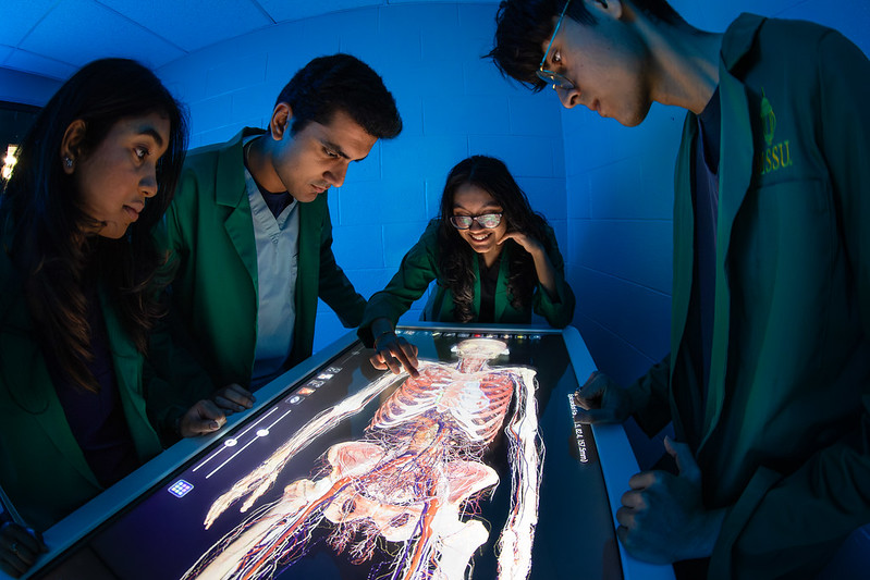
A costly expenditure, the Anatomage Table has many benefits: it allows up to 8 students at a time to view minute anatomical details without interacting with a real human body, and helps students to see the effects of various pathological variations on humans. Dr. Alla Barry, a professor of Anatomy and Physiology, Pathophysiology, and Advanced Human Dissection at MSSU, has explored the various functions of the table, and comments that it “provides an immersive and interactive learning experience for students and medical professionals. Users can explore anatomical structures in 3D, dissect virtual cadavers, and visualize complex anatomical relationships dynamically, enhancing understanding and retention.” She also says that “the Anatomage Table accurately represents human anatomy, [which] allows for a deeper understanding of anatomical structures [and] is particularly valuable for medical education and
surgical planning.” As an educator, she finds it extremely helpful that professors can “create custom presentations and case studies using the Anatomage Table, tailoring content to specific learning objectives or clinical scenarios.” The Anatomage Table also has important uses for clinical practice, as students can “simulate surgical procedures, pathology cases, and medical imaging techniques such as CT scans and MRI scans, [allowing] them to practice skills in a safe and controlled environment before working with real patients.” A last benefit Dr. Barry finds is that “some versions of the Anatomage Table support remote access, allowing students to engage with the content from anywhere with an internet connection.” As virtual learning becomes more prevalent, she comments that “this feature is particularly valuable in situations where students cannot physically access the Table.”
Few students have first handedly interacted with the cadaver table as of now, but one pre-medical student who was able to take a glimpse into the uses it offered was interviewed. When asked about a unique feature that the table offers, Aadhya Subhash, a second-year student in the MKEAP program at MSSU, said it has the “ability to produce a life-size image or visual image of the human body”. Furthermore, she finds it helpful to students’ understanding of anatomy because it allows for students to “highlight specific nerves or muscles or parts of the bones of the body”. Aadhya also mentioned that, alongside the real human cadavers in the lab, the table gives “a 3-D outlook on how certain nerves or bones in the body may appear as opposed to just viewing it through a two-dimensional textbook or image”, and allows for easy
digestion and understanding of course material. Lastly, she likes that students can emphasize and focus on specific areas of the body.
With the new Health Sciences Innovation Center on the way and other science and health resources being added to the school, MSSU is taking strides in providing opportunities for pre-health
students. The addition of the digital anatomical table to MSSU’s renowned cadaver lab will be greatly helpful when paired with cadavers and other unique health resources and facilities.


